
Authors: Syed Rahil / Editor: Lynn Stevenson, Liz Herrieven / Codes: / Published: 15/11/2022
Case scenario

Image 1 via www.dermnetnz.org
You are an Emergency Medicine registrar seeing a worried 35-year-old woman who presents to the Emergency Department (ED) with a history of rash on her upper limbs for the past 1 week. The rash started on her palms and has gradually spread proximally. It is very itchy, and it is affecting her sleep and her work.
There is no involvement of other body parts or mucous membranes. She was unwell and feverish for few days at the onset of the rash but has no other specific symptoms.
She does not have any previous medical problems, except an episode of urinary tract infection 2 weeks ago, which was treated with Nitrofurantoin. She is not taking any regular medications and she has no history of drug allergy.
What is the diagnosis and the management of this condition?
Introduction
The skin is the largest organ in the body. There are hundreds of skin diseases, many present with (often similar looking) rashes. Diagnosis of skin conditions can be a challenging task for non-dermatologists.
Like most investigations, skin specific investigations e.g. dermatopathology and biopsy, are done to ratify the clinicians provisional diagnosis. The key to which is history, examination and communication of examination findings.
History
Key questions include: the time of onset, duration, location, evolution, and symptoms of the rash or lesion.
Additional information on family history, occupational exposures, comorbidities, medications, and social or psychological factors is extremely helpful.
In suspected drug induced rash, detailed history of medications (including over the counter drugs) is important. Some hypersensitivity reactions may appear up to 3 months after a medication is taken.
Physical examination
The patient should always be examined in a good light and, if possible, with a magnifying lens. The physical examination includes visual inspection and palpation of the skin. The morphology, arrangement and distribution of the lesions are essential to note. In addition, colour, consistency and the number of lesions presented should be recorded.
Communication
Discussing a skin lesion with a dermatologist is similar to discussing an abnormal ECG with a cardiologist, the abnormality requires correct description in order to receive appropriate advice. Common dermatological terms and examples are listed below.
Lesion:
A single area of altered skin. It may be solitary or multiple.
Rash:
A widespread eruption of lesions.
Dermatosis:
Generic term for a disease of the skin.
How to describe a skin lesion based on morphology
Flat lesions
Macule:
Less than 1cm in diameter.

Patch:
More than 1cm in diameter.

Patch of caf au lait spot as seen in patients with neurofibromatosis.
Raised lesion
Papule:
Any solid lesion less than 1cm in diameter and that is raised above the surface.

Dermal melanocytic naevus (type of mole or naevus).
Nodule:
Any lesion more than 1 cm in diameter that is palpable between the finger and thumb.

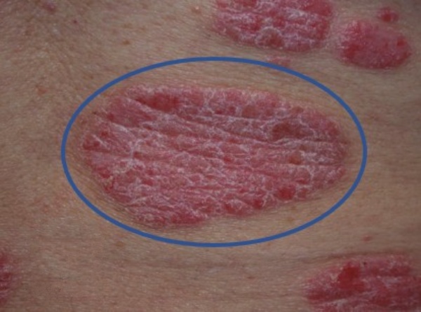
Plaque:
Any lesion more than 1cm in diameter where the diameter is > than the thickness i.e. the lesion can be felt only with fingertips.
Fluid filled lesion
Vesicle:
Fluid filled lesion that is <1cm in diameter.

Bulla:
A fluid filled lesion larger than >1cm in diameter.

Pustule:
Small skin lesions containing pus are defined as pustule and larger lesions as abscess.

Lesion due to broken skin
Erosion:
Loss of epidermis only is defined as erosion. Skin is made up of 3 layers, epidermis, dermis and subcutaneous tissue.

Ulcer:
Ulcer involves loss of epidermis and dermis.

Non-blanching rash (purpura)
Purpura is the name given to the discolouration of the skin or mucous membranes due to haemorrhage from small blood vessels.
Petechiae:
Small, purpuric lesions up to 2mm across.

Ecchymoses:
Larger areas of extravasations of blood. Also called bruise.

Palpable purpura:
Palpable purpura is purpura that can be felt. It is secondary to inflammation of the blood vessels (vasculitis).

(Note: systemic vasculitis has to be excluded before diagnosis of cutaneous vasculitis can be made).
How to describe a rash by arrangement and distribution
The location of one or multiple skin lesions and the arrangement of multiple lesions in relation to each other can suggest a particular diagnosis.
Common arrangements of lesions are detailed below.
Clustered:
As seen in herpes simplex infections.

Grouped:
As seen in dermatitis herpetiformis.

In dermatitis herpetiformis skin lesions are present in groups and usually present symmetrically over elbows, knees and buttocks.
Linear:
As seen in insect bites.

A linear distribution of rash is usually seen in insect bites and other conditions like contact dermatitis.
Zosteriform:
Band like unilateral skin lesions as seen in herpes zoster infection.

Coalescing or confluent:

A coalescing rash is where individual lesions merge together (as seen in this picture of acute urticaria).
Case scenario description and diagnosis
The skin lesions in the above-mentioned case scenario can be described as multiple discrete erythematous targetoid (target like) papules localised to upper extremities.
This rash is typical of erythema multiforme.
Erythema multiforme is an immune mediated condition affecting skin and mucous membranes. The most common trigger is the Herpes simplex virus. Other causes include medications including antibiotics like erythromycin, nitrofurantoin (in the case discussed above), penicillins, sulfonamides, and tetracyclines.
Erythema multiforme is classified into minor and major variants. In erythema multiforme major there is significant involvement of mucous membranes.
Diagnosis is mainly based on history and clinical examination. The rash typically starts in acral distribution (hands/ feet) and gradually spread centrally. Early lesions present as round, erythematous itchy papules, which later develop into characteristic target lesions.
Target lesions consist of three concentric rings. There is a central, dusky area of epidermal necrosis surrounded by a lighter oedematous area and then an erythematous margin peripherally.
Erythema multiforme is a self-limiting condition and most people recover within 4-6 weeks. Cutaneous lesions resolve without scarring.
Complications:
- Mucous membrane involvement can be painful and significantly limit oral intake.
- Ocular involvement can lead to more serious complications including:
- Keratitis
- Conjunctival scarring
- Uveitis
- Permanent visual impairment
Urgent ophthalmology review should be sought in this situation. The decision for hospitalisation should be based upon the extent of cutaneous and mucosal involvement as well as underlying co-morbidities.
Treatment is supportive like anti histamines and topical steroids to help with itching. Even though the commonest cause is herpes simplex virus infection, antiviral treatment has not shown any significant benefit. Offending medication should be stopped and avoided in future.
Note:
This blog article is a summary of the knowledge I gained from resources like www.dermnetnz.com, www.uptodate.com and the text book Clinical examination and differential diagnosis of skin lesions by Dan Lipsker.
About the author:
I am Dr. Syed Rahil, an ED doctor with special interest in dermatology. My qualifications are MBBS, MRCP, MRCEM, FRCEM and MSc Dermatology. I was trained in Dermatology at St. Johns Institute of dermatology, based in Guys and St. Thomas hospital, London. I was fortunate to learn from the most eminent dermatologists in this country. I have also passed Specialty exit exam in dermatology and worked for few years in Dermatology departments in UK and India. Currently I am employed full time in ED and working towards CESR. syedsabr@googlemail.com
References
- Image 1 – Erythema multiforme. DermNet.
- Image 2 – Melanocytic naevus. DermNet.
- Image 3 – Caf-au-lait macule. DermNet.
- Image 4 – Skin coloured lumps and bumps. DermNet.
- Image 5 – Neurofibromatosis. DermNet.
- Image 6 – Psoriasis. DermNet.
- Image 7 – Vesicular hand dermatitis. DermNet.
- Image 8 – Bullous pemphigoid. DermNet.
- Image 9 – Generalised pustular psoriasis. DermNet.
- Image 10 – Erosive pustular dermatosis. DermNet.
- Image 11 – Pyoderma gangrenosum. DermNet.
- Image 12 – Meningococcal disease. DermNet.
- Image 13 – Bleeding and bruising. DermNet.
- Image 14 – Cutaneous vasculitis. DermNet.
- Image 15 – Herpes simplex. DermNet.
- Image 16 – Dermatitis herpetiformis. DermNet.
- Image 17 – Arthropod bites and stings. DermNet.
- Image 18 – Herpes zoster. DermNet.
- Image 19 – Urticaria an overview. DermNet.
- Hidajat C, Loi D. Drug-mediated rash: erythema multiforme versus Stevens-Johnson syndrome. BMJ Case Rep. 2014 Sep 22;2014:bcr2014205543. doi: 10.1136/bcr-2014-205543.