
Authors: Sahra Haji / Editor: Charlotte Davies / Codes: / Published: 15/06/2021
Editors Note: Please note, the femoral nerve should be below the second layer of cardboard, not above
Most of us are convinced that fascia iliac blocks for pain relief are fabulous. Models to teach and practice on can be expensive. Inspired by one of the ED registrars I work with, Dr Grace OConnell, I am going to show how to make a simple model for simulating going through the fascial layers at the hip. These models can be used to teach emergency medicine (or any) doctors how to do a fascia iliaca block. The aim is to increase confidence and know-how for doing such procedures. And its always fun to do a bit of arts and crafts to add a bit of creativity to learning.
Learning objectives for teaching session:
– Landmark technique of fascia iliaca block for pain relief in patients with fractured neck of femur
– How to avoid erroneous intravascular injection of local anaesthetic
– Contents of femoral sheath
Step 1 – Start with the raw ingredients
- Sponge wipes (ours are from Tesco), can be different colours for different muscle layers
- Cardboard, thick enough for that pop pop sensation of going through fascia lata and fascia iliaca
- Scissors & glue
- Coloured straws to represent the neurovascular bundle, can be different sizes
- Cling film (optional but wraps everything up nicely) at the end

Step 2 – layer like a cake
- Use different-coloured sponges and place the fascia cardboard in between the sponges
- Note that 2 sponges are used in the middle layer to help separate out the femoral nerve straw from the femoral sheath contents, which are femoral artery and vein

Step 3 – make the layers fit
- Draw out a line on the cardboard, going around the sponges
- Cut out the cardboard so they take on the shape of the sponges
- Do not stick anything together just yet

Step 4 – find some straws or any tubes
- Use 3 different colours of straws to represent femoral nerve (green), femoral artery (red) and femoral vein (blue)
- The colours and sizes of the straws should be as close as possible to real-life colours of the femoral neurovascular bundle

Step 5 – layer the straws
- Place the straws in the middle-sponge layer in correct anatomical order from lateral to medial, with artery and vein in femoral sheath separate from the nerve

Step 6 – Everything comes together
- Carefully cut out inside of middle-layer sponge (pink) so that the straws sit snuggly within the sponges
- Glue everything together as shown on the right
- This step can be modified to add the ASIS and pubic tubercle demonstrated by the black circles using any round semi-circular object or by curving the cardboard

Step 7 – Prep your landmark
- Split diagonal imaginary line across sponges into thirds using your fingers


Step 8 – palpate the pulse
- Simulate palpating the pulse to ensure local anaesthetic is not injected into blood vessels with the models
- The straws can be attached to pumps containing ribena so that blood is passing through the straws that represent blood vessels
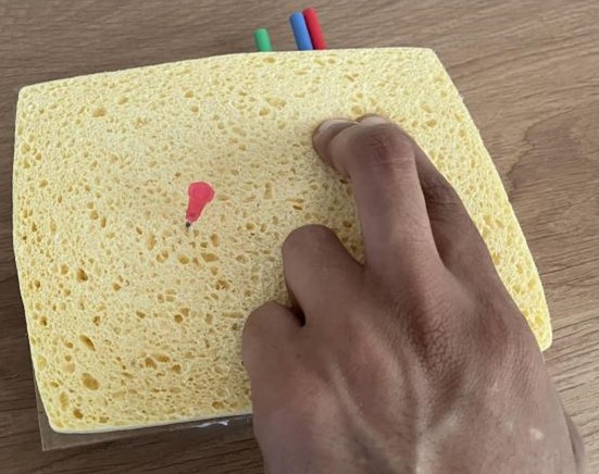
Step 9 – find your landmark
- Blunt needle can be inserted 2cm below the point at lateral 2/3 of diagonal line (away from blood vessels) – this is the injection point for fascia iliaca block

Step 10 – add some skin
- Wrap everything in cling film – this is the skin.

Step 11 – simulate block on a mannequin
- FIB model can be placed on mannequins hip area during simulation training
- More straws can be added when making the model to represent things like lymphatics
- Straw colours and sizes can be changed
- If you fill up a syringe with water / local anaesthetic and simulate doing the block and injecting, the liquid simply spreads and is absorbed by the sponge

- With the mannequin used here in our centre (above), piercing into the mannequin with the blunt needle through all the layers beyond the model prevents the easy flow of liquid being injected, which simulates the difficulty and obstructive sensation of injecting the local anaesthetic into the iliacus muscle during the block


Hopefully this has inspired you to make your own models for more teaching. Share your models using #rcemDIY or in the comments below and we’ll add as many pictures and images as we can. Here’s some starters:
First run at using wearable chest drain insertion model seemed to go okay. Hopefully adds a bit more patient communication to the skill. pic.twitter.com/EiimLuxwnG
Simon McCormick (@DrSimonMc) October 9, 2017
SPAM for USS guided cannulation…verdict was it’s better than nothing!
On to the next plan! pic.twitter.com/zmqQs0CBTwCharlotte Davies (@OneLongPlait) May 5, 2020

We don’t know where we found this awesome poster on simulation of corneal rust rings but we’d like to seek forgiveness for sharing, so get in touch!
There’s lots of suggestions for c-section models, placentas, and adaptations on twitter.

Mr Cardboard box is a regular patient in our ED for low fidelity sim around answering emergency buzzers, knowing where equipment is, systematic approaches – and even teamwork and human factors.


Haematoma blocks are hard to simulate but I’ve found a dog treat snapped, then taped to something rigid allows you to practice feeling for a fracture, and feeling for a crunch as you inject. To practice your colles reduction, there’s a simulator here.
For lateral canthotomy, have a look at the blog here, and then look at the simulator below.
How to make a Low Tech – Cheap Lateral Canthotomy model
This cheap model, can be used to practice the uncommon emergency. pic.twitter.com/brU2GVjIvK
Sarah E (@drsarahedwards) March 13, 2021
You can now complete RCEMLearning SBA on FIBs. Please log in to access it.
References and Further Reading
- The Royal College of Emergency Medicine, Best Practice Guideline. Fascia Iliaca Block in the Emergency Department. Revised: July 2020.
- RCEMLearning, Fascia Iliaca Block. 20123.
- Yang L, Li M, et al. Fascia iliaca compartment block versus no block for pain control after lower limb surgery: A meta-analysis. Journal of Pain Research Volume 10:2833-2841. 2017. Figure 1.
- Neill A, Anatomy for Emergency Medicine 028: Fascia Iliaca Block. Vodcast. Emergency Medicine Ireland. 2014.
- RCEMLearning, Simulation Overview. 2019.
- Images: Dr Sahra Haji, Dr Toni Robinson