Author: Charlotte Durand / Questions: Joshua Clough / Reviewer: Faathima Musaamil / Codes: / Published: 12/09/2018
Editor’s Note: Rashes are difficult to diagnose in both children and adults. The key is to be able to identify the important types and then manage to learn about the rest slowly. If a child is otherwise well – you’ll send them home with advice. If a child is poorly – they’re being admitted. The cause of the rash? This can be tricky sometimes. What is important is being aware of what ‘not to miss’: non-blanching rashes and possibilities of non-accidental injury. Most common presentations of rash are most likely associated with a virus, as after all, measles, rubella, chicken pox, etc. are all viral. Safety net carefully. I quite like PCDS websites as it gives you a list of differentials based on site symptoms or morphology – really useful!
As a Paediatric (Emergency Department) ED consultant I get asked many times a day “just to come and check a rash for me”. Often these will be innocent common rashes and we can reassure the parents and discharge the family home. Other times we won’t know what the rash is, but if we know the common rashes, then this gives us a fighting chance of being able to give the correct diagnosis.
At the other end of the spectrum it is vital to be aware of the more serious rashes which can indicate severe illness in babies and children.
I am going to go through some typical cases of paediatric rashes (with pictures) with the aim of giving you a good knowledge base when seeing these patients. The first section is about neonates and the second section looks at the “classic” rashes seen in older babies and children.
Part 1 – Neonates
I think its safe to say that most ED doctors have a slight tachycardia when faced with newborn babies with spots. They are usually accompanied by anxious (and exhausted) new parents who may or may not have done a google search and have now come to you with their worries!
Case 1
To start, you are asked to see a 3 week old baby with spots on their face, trunk, arms and legs. The spots have red bases with yellow pustular centres. The baby is feeding well and parents have no other concerns.
Images: used with gratitude from www.dermnetnz.org with use from creative commons
This is toxic erythema of the newborn. It occurs in 30-70% of full-term infants, and is therefore the most common pustular eruption in newborns. The aetiology is unknown. There are multiple yellow or white erythematous macules and papules (1-3mm in diameter) which can rapidly progress to pustules on an erythematous base. The lesions are distributed over the trunk and proximal extremities, but spare the palms and soles. It may be present at birth, but usually appears within 24-48 hours. Lesions typically resolve in 5-7 days, but may last several weeks.
Diagnosis is typically clinical, but can be confirmed by microscopic evaluation – numerous eosinophils. No treatment is needed.
Case 2
You are asked to see a 2 week old term baby who is otherwise well. Parents are concerned about these white spots on their baby’s nose.
These 1-2mm whitish yellow papules that are found on the nose, cheeks, chin and forehead are Milia.
These typically resolve in the first few weeks of life. They are secondary to the retention of keratin and sebaceous material in the pilacious follicles. These lesions disappear spontaneously.
Milia in newborns may also occur on the hard palate (Bohn’s nodules) or on the gum margins (Epstein’s pearls) as seen on the five week infant in the image above.
Case 3
You are asked to see a 2 day old baby as mum has noticed blisters on her lips and forearms. The baby is feeding well and there are no other concerns.
This is a diagnosis of exclusion – these are sucking blisters, i.e. oval, thick-walled vesicles or bullae that are filled with sterile fluid. Lesions may have erosion or crusting present. They are unilateral or bilateral and are usually located on the dorsal aspect of the wrists, hands, or fingers of neonates who are noted to suck excessively at the involved regions.
The mother of the baby also comments that the lesion keeps going a funny colour when on it’s side but then is back to normal within 5 minutes. The baby seems otherwise well. This is classical of a Harlequin colour change, observed when an infant is lying on their side, characterized by intense reddening of the dependent side and blanching of the non-dependent side, with a demarcation line along the midline. Lasts seconds to 20 minutes and resolves with increased muscle activity or crying. It affects 10% of newborns, and is entirely benign. Aetiology unknown.
Case 4
You are asked to see a 2 week old baby as the parents are concerned the baby has a large bruise on his lower back, this was not previously noted on the baby check. The mother is Chinese and dad is Caucasian.
Image: used with gratitude from www.dermnetnz.org with use from creative commons
Congenital dermal melanocytosis (previously known as the Mongolian blue spot) is the most frequently encountered pigmented lesion in newborns. It is seen more commonly in Asian, African American, and Hispanic neonates compared to Caucasian babies.
These congenital blue-grey pigmented macules with undefined borders can have a diameter of 10cm or more. Lesions are most commonly found in the sacro-gluteal region, or in the shoulders. They are completely benign lesions and usually fade during the first or second year of life. Mongolian spots can sometimes be difficult to differentiate from bruises and if there are concerns these may be bruises in a non-ambulant baby then senior advice should be sought for consideration of non-accidental injury or a bleeding disorder.
Case 5
A 2 week old baby, mum noticed a few spots on the baby’s face the previous day. The baby appears ok but mum reports him to be more quiet than usual. Dad has had a recent cold core. When you examine the baby, they have a few vesicular looking spots on their trunk, a temperature of 35.8 and appear very quiet when examined.
Image: used with gratitude from www.dermnetnz.org with use from creative commons
Neonatal herpes simplex virus is usually transmitted during delivery or through transplacental transmission of the virus. Contact with family members with cold sores also accounts for some cases, so it is worth asking specifically about family contact with cold sores even a few weeks ago (as incubation can be as long as 6 weeks). HSV 2 causes more cases than HSV 1.
It generally presents in weeks 1-2 of life but may be later. Neonates may present with local or disseminated disease. Skin vesicles are common with either type, occurring in about 55% overall. Other signs are often non-specific but include temperature instability, lethargy, hypotonia, respiratory distress, apnea, and seizures. Neonates without skin vesicles usually present with localised CNS disease. In neonates with isolated skin or mucosal disease, progressive or more serious forms of disease frequently follow within 7 to 10 days if untreated.
Incidence estimates range from 1/3,000 to 1/20,000 births. Diagnosis is by viral PCR of vesicular fluid (or CSF), immunofluorescence, or electron microscopy. Treatment is with high-dose parenteral acyclovir and supportive care. It has a high mortality and significant morbidity, but IV Acyclovir decreases the mortality rate in CNS and disseminated disease by 50% and increased the percentage of children who develop normally from 35-50% to 80%. The mortality rate of untreated disseminated disease is 85%; among neonates with untreated encephalitis, it is about 50%. Without treatment, at least 65% of survivors of disseminated disease or encephalitis have severe neurologic sequelae. Death is uncommon in those with local disease but without treatment these will progress to disseminated disease or CNS disease that may not be recognised.
Part 2 – Older Babies and Children
Case 1
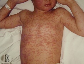
A 7-year-old boy, unwell for last few days with coryza, fever and conjunctivitis. Miserable and off food and drink. Up to date with immunisations.
Images: used with gratitude from www.dermnetnz.org with use from creative commons
Paramyxovirus (Measles)
Cases of measles tend to present in late Winter / Spring. Patient are infectious 1-2 days before prodrome to 4 days after onset of rash. The incubation period is 8-10 days, and normally asymptomatic until the 2-3 day prodrome of fever, malaise, anorexia, conjunctivitis, coryza, & cough. They may present with Enanthem – Koplik’s spots which are 1-3 mm whitish elevations with erythematous base, typically buccal mucosa opposite molar teeth.
The rash is a maculopapular, blanching rash that begins on the face, spreads cephalocaudally and centrifugally to involve the neck, upper trunk, lower trunk, and extremities.
The cough may persist for 1-2 weeks. Fever beyond the third or fourth day of rash suggests complication.
Complications of measles:
- Otitis media
- Bronchopneumonia
- Croup
- Encephalitis
- Myocarditis
- Pericarditis
- Subacute sclerosing panencephalitis (SSPE) years later
At risk:
- unvaccinated preschool-age children
- older children in whom vaccination failed ie if they have been vaccinated they can still get measles
The HPA suggest excluding a child from school/nursery for 4 days from the onset of rash. HPA provides recommendations for exclusion time for a range of childhood conditions.
Check out our podcast or reference guide.
Case 2
A 5-year-old girl, fever and unwell for last few days, has sandpaper type rash on trunk with an inflamed and red tongue.
Images: used with gratitude from www.dermnetnz.org with use from creative commons
This is scarlet fever, caused by Group A beta-haemolytic Streptococcus. Those at risk: <10 years (peak 4-8 years). It typically occurs from late Autumn to Spring, likely due to close contact indoors in school. The incubation period is 2-4 days. It presents with abrupt onset fever, headache, vomiting, malaise, sore throat. The infectious period is during the acute infection, and gradually diminishes over weeks. Northern Ireland and the HPA suggest excluding a child with scarlet fever from school until 24 hours after antibiotics started.
Examination findings include a bright red oral mucosa, palatal petechiae, tongue changes (strawberry tongue). The rash appears 12-48 hours after start of fever, usually starts below the ears, neck, chest, armpits & groin. It spreads to the rest of body over 24 hours and by about the 6thday of infection the rash starts to fade. Peeling, similar to sunburn, most prominent in the armpits, groin, fingertips (and/or toes). May continue up to 6 weeks.
Treatment is supportive, with antibiotics to reduce the infective period.
Purulent complications include otitis media, sinusitis, peritonsillar/retro-pharyngeal abscesses, Cervical adenitis. Non-suppurative sequelae include rheumatic fever, acute glomerulonephritis.
Case 3
A 4-year-old boy presents with a 5 day history of fever, spiking up to 40. They are miserable +++ with conjunctivitis, a red sore tongue, peeling fingertips, rash.
Creative Commons Attribution License Author: Dong Soo Kim
(A) Bilateral, non-exudative conjunctival injection with perilimbal sparing. (B) Strawberry tongue and bright red, swollen lips with vertical cracking and bleeding. (C) Erythematous rash involving perineum. (D) Erythema of the palms, which is often accompanied by painful, brawny edema of the dorsa of the hands. (E) Erythema of the soles, and swelling dorsa of the feet. (F) Desquamation of the fingers. (G) Erythema and induration at the site of a previous vaccination with Bacille Calmette-Gurin (BCG). (H) Perianal erythematous desquamation.
This is Kawasaki’s disease. Incomplete Kawasaki exists, but to formally diagnose complete Kawasaki Disease, you need 5 consecutive days of fever (usually >38.5 c, unremitting and not responsive to antipyretics) AND 4 of:
— Bilateral non-exudative conjunctivitis (90% have this)
— Erythema of the lips and oral mucosa
— Polymorphous rash
— Extremity changes (oedema and desquamation usually into second week of illness)
— Lymphadenopathy (usually anterior cervical nodes over SCM)
Some children may present with ‘incomplete’ Kawasaki disease, where there is less than 4 features present. These features may not be present at the time of your assessment. Therefore, you must specifically ask about these.
Blood tests must be taken, and characteristic lab results:
- CRP
- ESR
- Normocytic, normochromic anaemia
- Pyuria (urethral in origin)
Case 4
A 12-year-old unvaccinated boy from Eastern Europe is on holiday in the UK. He has been generally unwell with fever and nausea. A rash appeared today.
This is Rubella virus – at risk groups are unvaccinated adolescents. It often presents in Late Winter / early Spring. Incubation period: 14-21 days, Infectious period: 5-7d before the rash to 3-5d after the rash. Asymptomatic infection is up to 50%.
Prodrome in children is absent to mild. Adolescents & adults tend to have fever, malaise, sore throat, nausea, anorexia, painful occipital lymphadenopathy. Arthralgias/arthritis may be present in the older patients, and there is occasional peripheral neuritis, encephalitis, thrombocytopenic purpura (rare).
The rash is pinpoint, pink maculopapules that first appear on the face, spread caudally to trunk and extremities and become generalised within 24h.
Case 5
A 5-year-old, slightly unwell. Mum noticed a rash on her face this morning. She had a fever a few days ago but had seemed well.
Images: used with gratitude from www.dermnetnz.org with use from creative commons
This is slapped cheek/ erythema infectiosum caused by Human Parvovirus B19. School-age children are most at risk, sporadic outbreaks throughout the year. Incubation period: 4-14 days. Prodromal symptoms are non-specific e.g. fever, coryza, headache, nausea and diarrhoea. Infectious period: up until onset of the rash.
The rash can be very itchy, particularly on the soles of the feet. When occurring in adults (particularly women) can involve painful, swollen joints and malaise.
Pregnant women in contact with Parvovirus will likely need testing and follow up.
Case 6
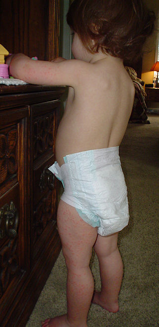
You spot this child with a rash, playing in the waiting room.

On closer inspection you see:

This is Roseola Infantum exanthem caused by Human Herpes Virus 6 (and 7). At risk: 6-36 months (peak age 6-7 months). Sporadic outbreaks throughout the year. Incubation period: 9 days. Prodrome: fever for 3-4 days, can be very high and is a common cause for febrile convulsions. Infectious period: asymptomatic persistent infection – virus intermittently shed into saliva throughout life. Abrupt defervescence with appearance of rash.
The rash is a blanching macular or maculopapular rash that starts on neck & trunk, spreads to face & extremities.
Case 7
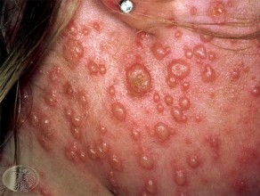
A pregnant mother brings in her 3 year old. He has chicken pox.
Images: used with gratitude from www.dermnetnz.org with use from creative commons
Varicella zoster virus (chicken pox) family herpes viridae. At risk: young children, sporadic outbreaks. Incubation period: 10-21 days. Prodrome: Ranges from asymptomatic to fever, malaise, cough, coryza and sore throat, spread via respiratory drop and vesicle fluid. Infectious period: 2 days before to approximately 5 days after onset of the rash, when the spots have scabbed over.
The rash has characteristic crops of macules, papules and vesicles.
Increased risk in adults, neonates & immunocompromised. Secondary bacterial infection in 5-10% – of these otitis media in 5%, pneumonitis, encephalitis , cerebellar ataxia (1:3000, lasts 2-3 months), hepatitis. Other complications include: Reye syndrome, Guillain-Barre, nephritis, carditis, arthritis, orchitis, uveitis.
The RCOG gives advice about pregnancy and chickenpox exposure.
Case 8
A 3-year-old with vesicular rash on chest, 3 spots in dermatomal distribution, low grade fever and rash seems painful but otherwise well.
Image: used with gratitude from www.dermnetnz.org with use from creative commons
Reactivation of latent VZV (shingles) in sensory ganglia. Elderly, immunocompromised and children who had chickenpox in utero or in first year of life. Prodrome is unusual in children.
Case 9
A 15-year-old boy with widespread maculopapular rash, sore throat and fever.
This is infectious mononucleosis (EBV) which presents with the triad of fever, tonsillar pharyngitis and lymphadenopathy, often with palatine pertechiae. Generalised maculopapular, urticarial or petechial rash occasionally seen. Can cause Gianotti-Crosti syndrome.
This itchy maculopapular or appears on extensor surfaces and pressure points 7 to 10 days after treatment with beta-lactams such as amoxicillin and cephalosporins. This rash indicates a hypersensitivity reaction to the antibiotic. It is not a true allergy and does not occur if the antibiotic is given later on in the absence of EBV infection.
Images: used with gratitude from www.dermnetnz.org with use from creative commons
Case 10
A 2-year-old with painful lesions in mouth (refusing to drink) also seems to have spots on hands and feet.
Images: used with gratitude from www.dermnetnz.org with use from creative commons
This is hand, foot and mouth disease due to coxsackie A16. It is highly contagious and preschool age children are at risk. Incubation period: 4-6 days. Prodrome (1-2 days before the rash) is fever, anorexia and a sore mouth. The spots can be very painful and tender to touch.
Case 11

Images: used with gratitude from www.dermnetnz.org with use from creative commons
Pityriasis Rosea is characterised by a large circular/oval “herald patch” on the chest, abdomen or back followed by smaller scaly patches in a Christmas tree distribution. Probably viral, HHV7 &8 implicated, not proven. It is seen in Spring, Autumn and winter affecting 10-35 year olds. There is a mild prodrome of malaise, nausea, anorexia, headache and low grade fever.
Case 12
Images: used with gratitude from www.dermnetnz.org with use from creative commons
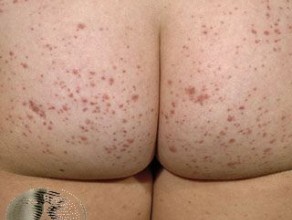
This is HenochSchnlein Purpura (HSP). The distribution of the rash is important- it’s normally on the bottom and on the back of the legs. If atypical distribution or the child is unwell, consider meningococcaemia, thrombocytopenia, or other rare vasculitides. HSP often presents with the “classic” triad of joint pain, abdominal pain and rash.
Joint Pain: Swelling and arthralgia of large joints are often the patient’s main complaint. Generally pain resolves spontaneously within 24-48 hours.
Abdominal pain: Uncomplicated abdominal pain often resolves spontaneously within 72 hours. Serious abdominal complications may occur including intussusception, bloody stools, haematemesis, spontaneous bowel perforation, and pancreatitis, so complete a thorough examination.
Renal Disease: Haematuria is present in 90% of cases, but only 5% are persistent or recurrent. Less common renal manifestations include proteinuria, nephrotic syndrome, isolated hypertension, renal insufficiency and renal failure (<1%). Renal involvement may only present during the convalescent period.
Subcutaneous oedema (scrotum, hands, feet, sacrum): This can be very painful.
Rare complications – pulmonary and CNS involvement.
Investigations should include: Urinalysis (for blood and/or protein), BP, consider bloods (FBC, U&Es, clotting). Generally supportive management but steroids can be considered.
Case 13
Next up is a 3 week old baby with a few spots on his face the previous day, now has peeling/raw areas in hands, groin and axillae with some lymphadenopathy. Seems sleepy and not waking for feeds.
Creative Commons Attribution License (by 4.0) Authors: OpenStax Microbiology Copyright Holders: OpenStax Microbiology
This baby needs admission for IV access and IV antibiotics as per local protocol for likely staphylococcal scalded skin syndrome.
Hopefully, this has given you a background of common neonatal rashes and the classical childhood exanthems and will mean the next time you pick up the card of the baby with a rash you will go and see them with a little more knowledge and confidence.


