Author: John Whittaker / Editor: Taj Hassan, Chris Wearmouth / Reviewer: Eugene Henry / Codes: A5 / Published: 25/10/2021
Context
Isolated facial muscle weakness is an uncommon presentation to the emergency department (ED) and may be quickly diagnosed by the unwary as Bells palsy.
The emergency clinician must be aware of two potential pitfalls when presented with a patient with facial weakness:
- Central (UMN) facial weakness must be differentiated from a peripheral (LMN) palsy
- A diagnosis of Bells palsy should only be made after the exclusion of other causes of a peripheral facial muscle weakness
Definition
Bells palsy is defined as an acute idiopathic peripheral (LMN) facial nerve paresis and is the most common cause of acute peripheral facial weakness [1].
To be able to diagnose Bells Palsy, there must not be other neurological deficit in the limbs or cerebellar signs, no parotid or neck masses must be present and there must not be any vesicles in the ear, nose or mouth present. Other differentials should always be considered e.g. Lymes disease, middle ear infection.
In the UK it has an incidence of approximately 20 cases per 100 000 person years [2], in other words around 50 cases per year will occur in an average ED catchment area.
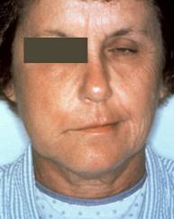
Right sided facial nerve palsy (Reproduced with permission from CDC)
Anatomy
Central Connections of the Facial Nuclei
An appreciation of the anatomy of the facial nerve is essential in understanding the clinical features and complications of facial paralysis.
The course and connections of the facial nerve are complex, but of particular clinical relevance to the EP are the central connections of the facial nuclei.
Each facial nucleus is bilaterally supplied by upper motor neurones from the cerebral cortex. Lesions within the brain therefore result in sparing of the muscles of the upper half of the face.
The image illustrates bilateral innervation of the upper half of the face which results in sparing of the forehead muscles in an upper motor neurone lesion.
Functional Anatomy of the Facial Nerve
Study the anatomy of the facial nerve illustrated below and then answer the question on the next page.
Note that the facial nerve runs through the internal acoustic meatus in the temporal bone close to the inner ear.
Pathophysiology
Herpes simplex virions
The cause of Bells palsy has long been debated but only recently has evidence started to accumulate for a viral origin.
Reactivation of latent herpes simplex or zoster virus is the most likely scenario, as shown in the image.
Herpes virus DNA has been isolated from facial nerve endoneural fluid [5], however attempts to isolate herpes simplex DNA from cerebrospinal fluid (CSF) and saliva in facial palsy patients, have been less successful [6,7].
History
Two questions must be asked by the clinician when assessing a patient who presents with an acute facial weakness:
- Is this an upper or lower motor neurone lesion?
- Are there any additional features that exclude Bells Palsy (idiopathic lower motor neuron facial nerve palsy)?
The most common presenting complaint in acute facial palsy is of unilateral facial weakness, which may have been noticed by the patient themselves or by a family member.
Although facial numbness is a commonly described in acute peripheral facial palsy, formal testing of the trigeminal nerve should demonstrate normal sensation. Subjective reduced sensation is caused by reduced facial muscle tone.
Depending on the cause of the facial nerve palsy, other features of the history may be elicited.
Approximately 50% of patients with Bells palsy experience facial pain, aural fullness or postauricular pain and in 25% of cases this precedes the paresis by 2-3 days [4].
Other symptoms, such as recent trauma, fever, headache, earache, rash, weight loss or other neurological or systemic upset may suggest an alternative cause other than Bells palsy.
Facial palsy (Reproduced with permission from Wellcome Images)
Examination
Is this an upper or lower motor neurone lesion?
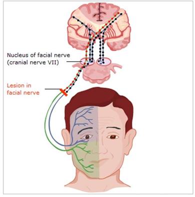
The key to differentiating a central (upper motor neurone) from a peripheral (lower motor neurone) facial palsy is to identify the extent of facial muscle weakness. The muscles of the upper half of the face (frontalis, corrugator and orbicularis) are innervated bilaterally by corticobulbar fibres. Weakness of the forehead muscles indicates a peripheral facial nerve problem whereas sparing of the forehead muscles is diagnostic of a central lesion. Asking the patient to close their eyes tightly or wrinkle their forehead will quickly identify the source of the problem.
Learning Bite
A patient with facial weakness who cannot close their eye tightly or raise their eyebrow has a peripheral facial palsy.
A lesion in the facial nerve resulting in weakness of all the facial muscles
Is this an idiopathic peripheral facial muscle weakness (Bells palsy) or is there another cause for the problem ?
Having identified a peripheral facial muscle weakness, the clinician must then exclude other causes before diagnosing an idiopathic palsy (Bells palsy).
General examination
Erythema migrans in a woman with Lyme disease (Reproduced with permission from CDC)
General examination of the patient should be normal in Bells palsy. Derangement of vital signs, fever, rash or other abnormality on examination all point to an alternative cause for facial weakness.
Causes of LMN facial nerve palsy with systemic upset include:
- Lyme disease
- Sarcoidosis
- HIV infection
In addition, acute peripheral facial weakness may either be found in known diabetics or be a presenting feature of previously undiagnosed diabetes [8].
Mononeuropathy in diabetes is most commonly caused by vascular occlusion and therefore has a poorer prognosis than facial weakness from other causes.
Head and Facial examination
Tumour of the parotid gland (reproduced with permission from Wellcome Images)
It is important to examine the face and head carefully as most of the alternate causes for peripheral facial weakness arise from involvement of the facial nerve in its extracranial course.
The commonest cause of peripheral facial weakness is Bells palsy but a careful history and examination must be performed to exclude other rarer causes.
Ear, nose and throat
Examine for signs of ipsilateral vesicular rash in/behind the ear, on lateral neck, on the tongue, buccal mucosa, tongue or palate, which may indicate Ramsey Hunt Syndrome.
Examine the ear for evidence of otitis media, cholesteatoma, or malignant otitis externa (spread of infection from the ear canal eroding into the skull base, most commonly seen in immunocompromised and elderly diabetics). The mastoid may also appear inflamed as a consequence of chronic ear infection. Any evidence of the above requires urgent ENT referral.
Careful examination of the parotid gland may reveal swelling, suggesting parotitis or tumour, compressing the facial nerve as it passes between the deep and superficial lobes of the gland.
Cranial nerves
Altered sensation, such as heaviness or numbness, is commonly described by patients with facial weakness and is thought to be due to reduced muscle tone. Formal sensory testing of the trigeminal nerve should prove to be normal.
Strictly speaking, Bells palsy is an isolated facial nerve paresis, but there is some evidence that other cranial nerves, particularly trigeminal, glossopharyngeal and hypoglossal may, very occasionally, be involved [9].
If any cranial nerve deficits are identified in addition to the facial nerve, it must not be classified as Bells Palsy, and the patient should undergo further investigation.
Trauma
Post traumatic facial nerve paralysis is most commonly caused by fracture of the temporal bone and may be immediate or delayed [10].
If delayed, the weakness appears, on average, 4-5 days after the injury and is caused by oedema, delayed arterial spasm or external compression by haematoma.
History of concerning trauma is an indication for imaging and discussion with ENT/neurosurgery.
Learning bite
Important differential diagnosis for a facial palsy, other than Bells Palsy, include:
- (Central) UMN causes, such as a stroke, SDH, or tumour
- (Peripheral) LMN causes
- Infective acute otitis media, cholesteatoma, malignant otitis externa, Lyme Disease, viral infection (including HSV-1, CMV, and EBV)
- Parotid pathology parotitis, malignancy
- Trauma or iatrogenic
- Neurological (Multiple Sclerosis , Guillain-Barr syndrome, Diabetic neuropathy, Myopathy etc)
- Vascular Vertebrobasilar insufficiency
As Bells palsy is an idiopathic condition, there is no diagnostic test currently available that will confirm the diagnosis. Although the aetiology is felt to be related to herpes virus infection, serum tests for rising antibody titres to herpes simplex are not reliable [11].
Serological testing will be required if other causes of peripheral facial weakness, such as Lyme disease or HIV, are suspected.
The only other investigations required are those to exclude a secondary cause for facial weakness. All patients must have their capillary blood glucose measured and routine observations performed.
In the presence of either a central facial weakness or a peripheral weakness with other features (e.g. history of trauma, systemic upset or co-existing neurological deficit), radiological imaging will be required.
Grading Severity
As facial muscle weakness can vary from mild to complete, grading it can help in the prognosis and follow-up. The modified House-Brackmann classification is the most commonly used scale as in the table below.
Table 1: The modified House-Brackmann classification
| Grade | Description | Characteristics |
| I | Normal | Normal facial function |
| II | Mild dysfunction | Slight weakness noticeable on close inspection. May have slight synkinesis |
| III | Moderate dysfunction | Obvious, but not disfiguring, difference between two sides. Compete eye closure with effort. Noticeable but not severe synkinesis, contracture or hemifacial spasm |
| IV | Moderately severe dysfunction | Obvious weakness or disfiguring asymmetry. Normal symmetry or tone at rest. Incomplete eye closure |
| V | Severe dysfunction | Only barely perceptible motion. Asymmetry at rest |
| VI | Total paralysis | No movement |
General Management Principles
Bells palsy can only be diagnosed when there is peripheral facial muscle weakness in the absence of an apparent cause. If any alternative features/causes have been identified, management should be directed there.
Management of facial nerve palsy can be divided into:
- Treatment directed at the cause of the facial nerve palsy
- Treatment of the consequences of the facial nerve palsy
Eye care
The most common problem associated with facial muscle weakness is incomplete closure of the eyelid. This may lead to exposure keratitis and corneal ulcers.
Two strategies should therefore be employed:
- Hourly lubricating eye drops during the day and eye ointment at night
- Eye taping at night
In addition, patients must be told to report new eye symptoms, such as pain, discharge and change in vision.
Patients with incomplete eye closure should be referred for ophthalmology follow-up as further options, including temporary tarsorrhaphy or botulinum toxin tarsorrhaphy, are available.
Learning bite
Eye care is extremely important in the management of Bells palsy as ophthalmological complications are common.
Attempts have been made to reduce facial muscle weakness with physiotherapy, electrical stimulation and acupuncture. None have high quality evidence to support their routine use [17, 18, 19].
Bells Palsy management
Treatment directed at the facial nerve has been a controversial area for many years, but recent high quality studies have clearly established the benefits of steroids in Bells palsy.
Steroids
Two recent large, high quality studies [12,13], and subsequent systematic reviews and meta-analyses [14,15], have all shown a reduction in time to complete recovery and an increased chance of complete recovery at three and nine months in patients treated with steroids (NNT 6 (95% CI, 4 to 9) for complete recovery at three months).
Both studies were restricted to patients with Bells palsy who presented within 72 hours; and in both studies they used prednisolone at a dose of 50 mg or 60 mg for a total of 10 days; one of the studies tapering the dose after 5 days [12,13].
Subgroup analysis from both systematic reviews and meta-analysis suggests that steroids may also be associated with improved recovery, even when started more than three days after the onset of symptoms [15].
NICE Recommends for people presenting within 72 hours of the onset of symptoms, consider prescribing prednisolone
Giving 50 mg daily for 10 days or
Giving 60 mg daily for five days followed by a daily reduction in dose of 10 mg (for a total treatment time of 10 days) if a reducing dose is preferred.
Learning bite
Treatment of patients with Bells palsy with corticosteroids has been proved to have beneficial effects on recovery time and to increase the chances of a complete recovery.
Antiviral agents
The effects of antiviral agents on recovery were also studied in the previously mentioned trials and further systematic reviews.
No positive benefit was demonstrated with the use of antiviral agents either used alone or in combination with prednisolone.
A meta-analysis directly comparing the use of steroids, with and without antiviral agents, confirmed this conclusion [16].
Learning bite
The addition of an antiviral agent to steroid treatment in Bells palsy has not been shown to provide any additional benefit.
Bells Palsy: Prognosis and Follow-up
In 70% of cases of Bells palsy facial paralysis is complete and, if untreated, almost 75% of patients will recover fully with 84% recovering full or near normal function [4]. It usually develops in 72 hours and does not progress after 72 hours.
Common sequelae found in those that fail to recover are:
- Residual partial facial weakness
- Facial contracture
- Ageusia loss of taste function (of the tongue)
- Motor synkinesis involuntary muscle movement accompanying a voluntary movement, e.g. eye closure when smiling
- Autonomic synkinesis e.g. crocodile tears syndrome when lacrimation occurs with salivation
Learning bite
84% of patients with Bells Palsy will recover full or near normal function without any treatment. Most people with Bells palsy make a full recovery within 9 months.
Although the vast majority of patients will regain normal or near normal facial function, there are certain prognostic indicators that predict a poorer outcome [20]:
- Increased age
- Hypertension
- Impairment of taste
- Pain other than in the ear
- Complete facial weakness
Follow-up of a patient with Bells palsy who attends the ED should be arranged either through the GP or ENT clinic, according to local policy.
Recurrence of Bells palsy is very rare but, if a second episode does occur, then an alternative diagnosis should be considered, e.g. myasthenia gravis or brain stem lesion [20].
Ramsay Hunt Syndrome: Introduction
Ramsay Hunt syndrome is defined as a peripheral facial nerve weakness accompanied by a vesicular rash.
Cause
It is caused by herpes zoster infection of the facial nerve and geniculate ganglion, although the exact pathophysiological processes involved are still unclear [21].
Presentation
Ramsay Hunt Syndrome with a vesicular rash in the right ear (Reproduced with permission from Wellcome Images)
Presentation is with a classic peripheral facial nerve weakness, but close examination will reveal an ipsilateral rash that may be evident in one of a number of areas dependant on the sensory ganglion(s) involved:
- In the ear canal or behind the ear
- Over the lateral neck
- On the tongue or buccal mucosa
- On the palate, uvula and/or pharynx
It is important to note that the rash may be initially erythematous only, with vesicles developing later.
Associated symptoms
Ramsay Hunt syndrome is commonly accompanied by associated symptoms, such as hearing loss and vestibular disturbance due to involvement of structures adjacent to the facial nerve [4]. It is associated with a poorer prognosis than Bells palsy and sequelae, such as persistent synkinesis and hearing loss, are more common in Ramsay Hunt syndrome [22].
Ramsay Hunt Syndrome: Treatment
Although high quality studies are lacking, best evidence suggests that a combination of high dose steroids and aciclovir (800 mg five times a day for 7 days) are associated with improved rates of recovery [23,24].
Other treatment and follow-up for the sequelae of facial muscle weakness are also required, as for Bells palsy.
In addition, the patient must be counselled on the infectivity of the rash and potential risk to the non-immune, pregnant and immunocompromised.
Learning bite
It is important to examine a patient with apparent Bells palsy very carefully, as evidence of the vesicular rash may be difficult to see and management of the patient is therefore significantly altered.
Ramsay Hunt Syndrome: Zoster Sine Herpete
Bells Palsy (Reproduced with permission from Wellcome Images)
Zoster sine herpete is a term for Ramsay Hunt syndrome without apparent vesicles or when there is a delayed presentation of the rash. The image shows facial paralysis, post herpes zoster (reproduced with permission from Wellcome Images).
Learning bite
In 15% of patients with Ramsay Hunt syndrome, vesicles develop after the onset of facial weakness [4] and therefore patients diagnosed with Bells palsy must be instructed to return or see their GP should a rash develop later.
The following are potential pitfalls the unwary clinician may fall into when assessing and managing a patient with an acute facial palsy:
- Failure to differentiate an upper from a lower motor neurone lesion, thereby missing a potentially significant central lesion.
- Inadequate examination missing an alternate cause for a lower motor neurone palsy e.g. parotid gland pathology.
- Failure to prescribe early high dose steroids in a patient diagnosed with Bells palsy.
- Failure to manage the sequelae of facial palsy, in particular, adequate eye care.
- Failure to identify a rash and miss a diagnosis of Ramsay Hunt syndrome.
- Bells palsy is an acute idiopathic peripheral facial nerve paresis.
- if untreated, almost 75% of patients will recover fully with 84% recovering full or near normal function. (level of evidence 4)
- Intact forehead muscle function, revealed by an ability to raise the eyebrow or close the eye tightly, is a sign of a central (upper motor neurone) lesion.
- Bells palsy is the commonest cause of a peripheral facial muscle weakness, but a careful history and examination must be completed to exclude one of the many other causes of this problem. (level of evidence 5)
- The use of high dose steroids in Bells palsy has been shown to shorten recovery time and increase the chances of a complete recovery. (level of evidence 1a)
- Antiviral drugs do not have any beneficial effects on the time to recovery or incidence of complete recovery in Bells palsy. (level of evidence 1a)
- Eye care is extremely important in the management of patients with facial muscle weakness in the ED as ophthalmological complications are common in these patients. (level of evidence 5)
- Best evidence suggests that patients with Ramsay Hunt syndrome treated with a combination of high dose acyclovir and prednisolone have improved recovery rates. (level of evidence 4)
- Adour KK, Byl FM, Hilsinger RL Jr et al. The true nature of Bells palsy: analysis of 1,000 consecutive patients. Laryngoscope 1978;88:787-801. View abstract
- Rowlands S, Hooper R, Hughes R. et al. The epidemiology and treatment of Bells palsy in the UK. Eur J Neurol 2002;9:63-67. View abstract
- Bird TD. Nicolaus A. Friedreichs description of peripheral facial nerve paralysis in 1798. J Neurol Neurosurg Psychiatry 1979;42:56-58. View abstract
- Peitersen E. Bells palsy: the spontaneous course of 2,500 peripheral facial nerve palsies of different etiologies. Acta Otolaryngol Suppl 2002;549:4-30. View abstract
- Murakami S, Mizobuchi M, Nakashiro Y et al. Bell palsy and herpes simplex virus: identification of viral DNA in endoneural fluid and muscle. Ann Intern Med 1996;124:27-30. View abstract
- Kanerva M, Mannonen L, Piiparinen H et al. Search for herpesviruses in cerebrospinal fluid of facial palsy patients by PCR. Acta Otolaryngol 2007;127:775-779. View abstract
- Lazarini PR, Vianna MF, Alcantara MP et al. Herpes simplex virus in the saliva of peripheral Bells palsy patients. Braz J Otolaryngolog 2006;72:7-11. View abstract
- Saito O, Aoyagi M, Tojima H et al. Diagnosis and treatment for Bells palsy associated with diabets mellitus. Acta Otolaryngol Suppl 1994 511:153-155. View abstract
- Benatar M, Edlow J. The spectrum of cranial neuropathy in patients with Bells palsy. Arch Int Med 2004;164:2383-2385. View abstract
- Baumann BM, Jarecki, J. Posttraumatic delayed facial nerve palsy. Am J Emerg Med 2008;26:115.e1-2.
- Holland NJ, Weiner GM. Recent developments in Bells palsy. BMJ 2004:329;553-755. View abstract
- Engstrm M, Stjernquist-Desatnik A, Axelsson S et al. Prednisolone and valciclovir in Bells palsy: a randomised, double-blind, placebo-controlled, multicentre trial. Lancet Neurol 2008;7:993-1000.
- Sullivan FM, Swan IRC, Donnan PT et al. Early treatment with prednisolone or acyclovir in Bells palsy. N Eng J Med 2007;357:1598-1607. View abstract
- de Almeida JR, Al Khabori M, Guyatt GH et al. Combined corticosteroid and antiviral treatment for Bell palsy: a systematic review and meta-analysis. JAMA 2009;302:985-993. View abstract
- Goudakos JK, Markou KD. Corticosteroids vs corticosteroids plus antiviral agents in the treatment of Bell palsy: a systematic review and meta-analysis. Arch Otolaryngol Head Neck Surg 2009;135:558-564. View abstract
- Quant EC, Jeste SS, Muni RH et al. The benefit of steroids plus antivirals for treatment of Bells palsy: a meta-analysis. BMJ 2009;339:b3354. View abstract
- Beurskens CH, Heymans PG. Positive effects of mime therapy on sequelae of facial paralysis: stiffness, lip mobility, and social and physical aspects of facial disability. Otol Neurotol 2003;24:677-681. View abstract
- He L, Zhou MK, Zhou D, Wu B, Li N, et al. Acupuncture for Bells palsy. Cochrane Database Syst Rev. 2007 Oct 17;(4):CD002914.
- Best Evidence Topics (Buttress SJ, Herren K) [Internet]. Electrical stimulation and Bells palsy. BestBets; Manchester Royal Infirmary; c2002 View link
- Gilden DH. Bells palsy. N Eng J Med 2004;351:1323-1331. View abstract
- Sweeney CJ, Gilden DH. Ramsay Hunt syndrome. J Neurol Neurosurg Psychiatry 2001;71:149-154. View abstract
- Robillard RB, Hilsinger RL Jr, Adour KK. Ramsay Hunt facial paralysis: clinical analyses of 185 patients. Otolaryngol Head Neck Surg 1986;95:292-297. View abstract
- Murakami S, Hato N, Horiuchi J et al. Treatment of Ramsay Hunt syndrome with acyclovir-predisone: significance of early diagnosis and treatment. Ann Neurol 1997;41:353-357. View abstract
- Kinishi M, Amatsu M, Mohri M et al. Acyclovir improves recovery rate of facial nerve palsy in Ramsay Hunt syndrome. Auris Nasus Larynx 2001;28:223-226. View abstract
- NICE CKS Bells Palsy Management Scenarios. 2019.


